
3 records found...
-
-
The effects of low electromagnetic field and lead acetate combination on some hemato-biochemical and immunotoxicological parameters in mice
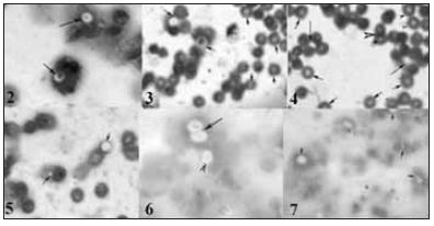
Figure 2. Phagocytosis of C. albicans by neutrophils of control mice (gp.1). One cell of C. albicans was engulfed by neutrophils (arrow), Leishman’s stain, X100. Figures 3-4. Phagocytosis of C. albicans by neutrophils of EMF exposed mice (gp.2). One and two C. albicans engulfed by neutrophils were represented by arrow (Fig. 2) and arrow-head (Fig. 3) respectively, Leishman’s stain, X100. Figures 5. Phagocytosis of C. albicans by neutrophils of mice administered lead acetate (gp.4). One C. albicans was engulfed by neutrophils (arrow), Leishman’s stain, X100. Figures 6. Phagocytosis of C. albicans by neutrophils of mice administered lead acetate (gp.5) showing two cells of C. albicans were engulfed by neutrophils (arrow) and one cell attached to the surface of neutrophil (arrow-head), Leishman’s stain, X100. Figures 7. Phagocytosis of C. albicans by neutrophils of mice administered lead acetate (gp.5) showing two cells of C. albicans were engulfed by neutrophils (A) and one cell attached to the surface of neutrophil (B) and/ or engulfed (arrow), Leishman’s stain, X100.
-
The effects of low electromagnetic field and lead acetate combination on some hemato-biochemical and immunotoxicological parameters in mice
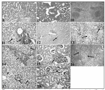
Figure 8-10. Liver, kidney and spleen sections from control mice (Gp. 1) showing normal structure respectively, H&E., x1200. Figure 11. The liver section from mice of (Gp. 2) showed focal centrolobular necrosis of the hepatic cells surrounded by severe hydropic degeneration involving the majority of hepatic parenchyma and round cells infiltration, H&E., x1200. Figure 12. The kidney section (Gp. 2) suffered from congestion of renal blood vessels, contracted glomerular tufts of some glomeruli and focal leukocyte aggregation, H&E., x300. Figure 13. Spleen section (Gp. 2) showed lymphoid depletion in splenic white pulps and hemosiderosis in red and white pulps, H&E., x1200. Figure 14. Liver section from mice given 5mg/kg BW (gp. 4) of lead acetate showed leukocyte infiltration in the portal area and hydropic degeneration, H&E., x1200. Figure 15. Kidney section from mice given 10mg/kg BW of lead acetate (Gp. 5) revealed focal replacement of renal parenchyma by lymphocytes, H&E., x1200. Figure 16. Spleen section from Gp. (5) showed severe lymphoid depletion, hemosiderosis in red and white pulps beside thickened splenic trabeculae, H&E., x1200. Figure 17. Liver section of mice exposed to a combination of lead and EMF (Gp. 6) showed focal replacement of hepatic parenchyma by lymphocytes, hyperplastic Kuffer cells, hydropic degeneration, hyperchromatic nuclei and disorganized hepatic cords, H&E., x1200. Figure 18. Kidney section from mice of Gp. (6) showed periglomerular lymphocytic infiltrations with individual coagulative necrosis, H&E., x1200.
Sayfa 1 / 1 -- Toplam Kayıt 3 -- Listelenen kayıt sayısı 3
0 records found...
Sayfa 1 / 1 -- Toplam Kayıt 0 -- Listelenen kayıt sayısı 0
|
 |
|
|
|
|